
Horizontal Fingernail Lines
A 78-year-old man with metastatic prostate cancer presented to his primary care physician with “white lines” that had appeared on his fingernails over the past year. He was asymptomatic and had no specific complaints regarding his nails. He denied any pain, breakage, or changes in nail growth.
He was diagnosed with prostate cancer initially 30 years before the current presentation. His disease remained stable until 3 years previous to the current visit, when metastases to the anorectal junction were discovered, necessitating initiation of chemotherapy. This started in June of 2021 and included Docetaxel, prednisone, Neulasta, Cabazitaxel, and leuprolide. His past dermatologic history included actinic keratosis and seborrheic keratosis. Additional medical history included hyperlipidemia, hypertension, impaired glucose tolerance, gastroesophageal reflux disease, gout, and asthma. His medications included allopurinol, amlodipine, ethacrynic acid, gabapentin, losartan, omeprazole, prednisone, pregabalin, albuterol. He had never smoked and usually consumed five alcoholic beverages per week. He was retired.
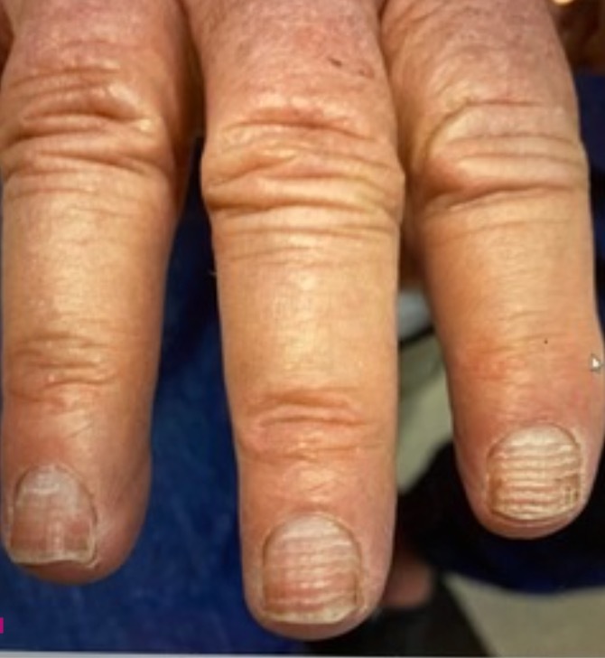
On closer examination, narrow, transverse, evenly spaced hypopigmented grooves were present on all ten fingernails (Figure 1). There was no cuticle involvement or skin changes surrounding the nails. The remainder of his physical examination was noncontributory.
Differential diagnosis:
- Mee's lines
- Melanonychia
- Muehrcke lines
- Beau's lines
- Leukonychia
- Traumatic nail dystrophy
Discussion
These horizontal lines represent Beau lines or Beau’s lines.
Beau’s lines are hypopigmented transverse grooves that can be visualized running horizontally across a fingernail or toenail. The nail will show a transverse depression that migrates distally as the nail continues to grow.1 This phenomenon results from temporary arrest of proximal nail matrix proliferation, which can be induced by cytotoxic chemotherapeutic agents such as Docetaxel.2 The nail growth is thinner during the cycles of chemotherapy, leaving a succession of horizontal grooves in the nails marking each round of chemotherapy. In Beau’s lines, the depth of the depression seen in the nail is directly related to the degree of damage, and width correlates to the duration.1 Because a fingernail takes approximately 40 days to emerge from the proximal nail fold, damage often does not become visible until 6-8 weeks after the insult.3 In the case of cytotoxic insult due to chemotherapy, the appearance of Beau’s lines can be correlated directly to the patient’s chemotherapy schedule.
Other possible etiologies for Beau’s lines include non-chemotherapeutic drugs such as retinoids, trauma to the nail, Raynaud’s disease, viral infections such as Coxsackievirus, Kawasaki disease, pemphigus vulgaris and local cutaneous disease such as paronychia or dermatitis. Because Beau’s lines represent a transient modification of nail plate morphology, no treatment is required other than addressing the underlying etiology. Some patients may choose over-the-counter supplements such as biotin or paint-on agents such as water-soluble nail solutions containing hyaluronic acid and Pistacia lentiscus to decrease risk of nail breakage and improve brittle appearance.4 However, caution should be exercised when taking over-the-counter biotin, as it may impact thyroid function testing.
Mee’s lines represent a change in the color of the nail without any change in nail thickness or palpable ridges, typically described as parallel white bands traversing the entire nail bed horizontally.2 Mee’s lines are indicative of arsenic, thallium, or other heavy metal poisoning; this patient did not possess any risk factors for such exposures.5
Melanonychia are vertical hyperpigmented lines that can represent a normal variant in the majority of patients, especially in people with darker skin tones. However, it must be differentiated from subungual melanoma clinically by identifying any sudden changes in the appearance of the bands, band width greater than 3 mm, involvement of the cuticle or nail fold (Hutchinson sign), or nail plate disruption.6 Vertical nail findings such as melanonychia are very uncommon in fair-skinned people and are a red flag for systemic disease.7
Muehrcke lines represent transverse white lines that appear in pairs and are not associated with nail thickness changes. They are caused by localized pathology within the nail bed, such as edema from hypoalbuminemia.5 Because they originate in the nail bed and not the nail plate, they do not migrate distally as the nail grows and they disappear when pressure is applied to the nail due to compression of the abnormal blood supply.7
Leukonychia represents an abnormal keratinization of the nail matrix, causing nonuniform hypopigmented changes to the nail plate.5 Although they can appear as horizontal white lines that migrate with nail growth, the changes can occur in a single nail or multiple, are rarely identical across nails, and is a common finding with no clinical significance.8
Traumatic nail dystrophy is a finding that arises due to repeated manipulation of the nail plate, as in frequent manicures and pedicures, scrubbing, picking, and biting, and is rarely uniform across all nails. Traumatic nail dystrophy typically presents as midline splitting or indentation, but can appear as a transverse splitting especially in the case of musicians or artists that use the index finger frequently when playing instruments or crafting.5 It can be accompanied by inflammation or thickening of the proximal nail fold and/or changes to the cuticle or nail plate.5
Treatment for nail abnormalities largely involves addressing underlying etiology. Information taken from references 1-8.
Data Sources
A PubMed search was conducted in Clinical Queries using key terms including Beau’s lines, Mee’s lines, horizontal nail changes, and nail abnormalities. This search included cohort studies, randomized controlled trials, meta-analyses, and reviews. Also searched were ClinicalKey and DynaMed. The search was conducted April 3, 2022.