BRIEF BACKGROUND
Extravasation of urine into retroperitoneal or peritoneal spaces can lead to fluid collections called urinomas. When infected, they progress to life-threatening sepsis, particularly in patients with impaired immune system or altered tissue healing. Also, retroperitoneal hematoma formation is a possible complication of anticoagulation,1,2 and when coupled with immunosuppression, may predispose patients to secondary complications like urinoma formation. Concurrent use of anticoagulation therapy, especially in those with a history of thromboembolic disease, further complicates management due to increased bleeding risk.1
Urinoma causes can be categorized into traumatic, obstructive, and spontaneous etiologies.3,4 Traumatic causes include blunt or penetrating injury to the kidneys, ureters, and bladder as well as iatrogenic injury after abdominal and pelvic surgeries or procedures. Obstructive causes include calculi, cancer, and congenital anomalies. Spontaneous urinoma formation is rare and has been reported in association with acute interstitial nephritis with nonsteroidal anti-inflammatory use.3,4
Pemphigus vulgaris (PV) is a severe chronic autoimmune blistering disease affecting the skin and mucous membranes.5 In this report, we discuss a case of a 70-year-old woman with PV on chronic steroids and a history of deep vein thrombosis (DVT) on anticoagulation, who developed a retroperitoneal urinoma secondary to ureteral necrosis. This ultimately led to sepsis, requiring extensive multidisciplinary intervention. This case highlights the challenges of balancing anticoagulation and immunosuppression in a medically complex patient, and the diagnostic considerations needed when managing retroperitoneal collections in such contexts.
CASE REPORT
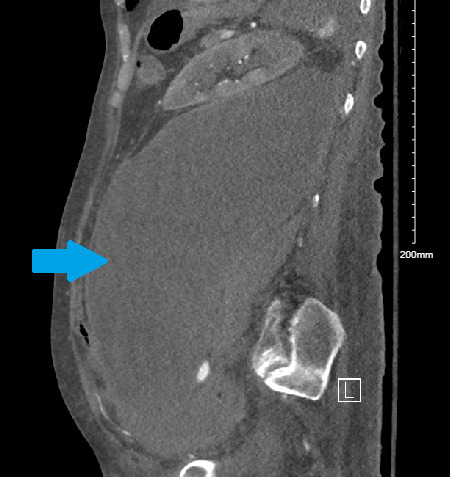
We present the case of a 70-year-old female with a history of PV on chronic steroid therapy and DVT status post inferior vena cava (IVC) filter placement on chronic anticoagulation with apixaban. She also had a chronic retroperitoneal hematoma. The patient presented with poor appetite and somnolence. In the emergency department, she was hypotensive with blood pressure of 70/50 mmHg and tachycardic with a heart rate of 110 bpm, reporting diffuse abdominal discomfort. She was afebrile, with clear lung sounds, and no abdominal tenderness on exam. Initial workup was significant for a lactate level of 3.6 mmol/l, which trended upwards to 4.2 mmol/L before fluids were administered, and a low white blood counts of 3.08 x10(9) /L (table 1). Her imaging revealed an increase in the size of her left retroperitoneal hematoma and perivascular inflammatory changes, raising concerns for a superimposed infection (figure 1). Sepsis treatment was initiated after which the patient was hemodynamically stable. However, a new left-sided hydronephrosis and concern for ureteral extravasation on imaging raised suspicion for urinoma as a source of sepsis. The hematoma size measured 26.4 x 12 x 10 cm on imaging. Radiologic evaluation demonstrated a 1.4 cm focus of contrast blush consistent with active urine extravasation but the urinoma was challenging to measure as imaging primarily visualized the contrast blush.
_showing_left_retro-peritoneal_fluid_collection_(blue_arr.png)
Interventional radiology was consulted. The patient underwent percutaneous nephrostomy tube placement to decompress the left kidney and retroperitoneal drain placement to drain the retroperitoneal fluid collection. Fluid studies confirmed accumulation of urine with heavy growth of proteus mirabilis in culture. Urology was then consulted and performed a left ureteroscopy, left ureteral stent placement, ureteral biopsy, and nephrostomy tube replacement for the left ureter. An atypical finding was noted of a portion of the left ureter being significantly damaged and necrotic. Contrast extravsation confirmed a ureteric fistula. The patient was managed with a bladder catheter to drain the bladder while other drains were maintained in order to allow the left ureter to heal. A tailored antibiotic plan was initiated based on sensitivities and guidance of the Infectious Disease team.
The patient’s hospital course was further complicated by the necessity to initiate anticoagulation given the extent of DVT while also requiring continued steroid therapy for PV control. Despite multiple anticoagulation regimens, the patient continued to have ongoing bleeding from the drains and worsening anemia which necessitated withholding anticoagulation at various points. Hematology was consulted and recommended a cautious approach of starting a prophylactic dose of anticoagulation with gradual escalation to therapeutic dosages as tolerated.
The patient was discharged back to the nursing home with a lower dose of anticoagulation and a tapering steroid course. Urology planned follow up imaging with antegrade nephrostograms to assess for timing for drain and stent removal. However, on a follow up outpatient visit with urology, she appeared fatigued and unwell. Due to concerning vitals, she was subsequently re-admitted due to worsening decubitus ulcer that has originally been noted in the first hospitalization.
DISCUSSION
This case highlights the complexities of managing a patient with multiple chronic conditions, including the interplay between chronic immunosuppression due to PV, anticoagulation due to an extensive DVT despite IVC filter, and the development of a retroperitoneal hematoma that turned into a urinoma and eventual sepsis. The combination of anticoagulation therapy and steroids increased the risk of bleeding due to the anticoagulation effects and the steroid-induced fragility of her blood vessels and tissues. This heightened bleeding risk likely contributed to the formation of the retroperitoneal hematoma, which subsequently led to the development of a urinoma due to compression and obstruction of the urinary tract by the retroperitoneal collection.1,2,6 Evidence demonstrates that glucocorticoids significantly increase bleeding risk when combined with non-vitamin K oral anticoagulants like apixaban.7,8
Temporarily holding anticoagulation therapy was critical in managing the patient’s bleeding risk and helped stabilize hemoglobin levels. This decision facilitated effective drainage and antibiotic therapy for sepsis, which is essential for improving patient’s outcomes.9 This collaborative effort of dermatology, hematology, and radiology along with primary team was crucial in managing the complex interplay of multiple organ systems and pathologies involved in this patient’s care.9–11
The American College of Cardiology recommends immediate discontinuation of anticoagulants and antiplatelet agents for patients with major bleeding, including retroperitoneal hemorrhage, combined with aggressive hemodynamic stabilization and early involvement of appropriate services such as surgery or interventional radiology.12 Retroperitoneal bleeding is specifically identified as a critical site bleeding that required definitive management by specialized services.12 Resumption of anticoagulation requires careful consideration for both thrombotic and bleeding risks through multidisciplinary consultation.12 For patients with high bleeding risk (relative or absolute contraindication to restarting anticoagulation) and high thrombotic risk, non-pharmacological therapies such as left atrial appendage closure devices for atrial fibrillation or retrievable inferior vena cava filters for acute venous thromboembolism may be considered, as was the case in our patient.1
Managing retroperitoneal hematomas presents significant clinical challenges due to diagnostic and therapeutic complexities.13 Diagnostic difficulties arise from the often nonspecific clinical presentation, which can delay recognition and diagnosis. Additionally, the lack of a clear consensus on optimal management complicates clinical decision-making, as treatment must be individualized based on patient’s stability ranging from conservative management, interventional radiology approaches, or surgical intervention.13
Retroperitoneal hematomas can cause pressure necrosis complications, especially when large or rapidly expanding, through compression of local structures and development of compartment syndrome.14–16 Femoral neuropathy occurs when hematomas compress the femoral nerve as it traverses the iliacus muscle.17 Bilateral ureteral obstruction and renal failure can result from direct compression of both ureters in the true pelvis, creating an intrapelvic compartment syndrome analogous to abdominal compartment syndrome.15
It is of note that urinomas are very rare complications that can lead to the presentation of an acute abdomen.18 Urinomas in literature can vary in size and range from a few centimeters to greater than 20cm.19,20 The size of urinoma depends on the duration of urine leak, the severity of distal obstruction, ongoing kidney function, and the ability of the perirenal fascia to confine the fluid.4
This case underscores the importance of a comprehensive evaluation of patients with complex histories and multiple comorbidities. It also highlights the challenges in diagnosing the source of sepsis and managing hemodynamic instability in such cases. Timely intervention, careful fluid management, and appropriate antibiotic therapy were critical in improving the patient’s condition. Weighing benefits and risks while keeping patient and medical power of attorney informed and active in the discussion was essential as well.
Acknowledgement
None of the authors has any financial or non-financial competing interests.