BRIEF BACKGROUND
Vitamin B12 (cobalamin) deficiency is common but underrecognized. In the United States, 3–4% of adults under 60 years are deficient, and the figure rises to 6% in those 60 years and older.1,2 Marginal depletion affects up to 20% of older adults.2 The serum cobalamin assay is unreliable in the 200–400 pg/mL range, where clinical decisions are hardest.3,4 Methylmalonic acid (MMA) resolves this ambiguity. Savage et al. demonstrated 98.4% sensitivity for clinically significant deficiency when MMA was the diagnostic standard.5 Despite this, MMA testing remains underutilized in primary care.4
Severe B12 deficiency can produce pseudo-thrombotic microangiopathy (pseudo-TMA). The syndrome mimics thrombotic thrombocytopenic purpura (TTP) closely enough that in one systematic review, 16 of 53 patients with pseudo-TMA received plasmapheresis before the correct diagnosis was made.6,7 Pernicious anemia clusters with organ-specific autoimmune diseases. Type 1 diabetes mellitus (T1DM) and vitiligo are the most common associations, yet no major society mandates routine cobalamin screening in these populations.8
GLP-1 receptor agonists, though not a factor in this patient’s case, place a growing outpatient population at risk for nutritional deficiency, and a 2025 joint advisory names B12 as a nutrient of concern during therapy.9
CASE REPORT
A 46-year-old man with T1DM (two years, insulin pump) and vitiligo presented with seven to eight days of intractable nausea and vomiting. Home medications included insulin via pump; the patient was not receiving a GLP-1 receptor agonist. Family history included lupus. Vital signs were stable.
Emergency department studies revealed severe pancytopenia: hemoglobin 6.5 g/dL (65 g/L), white blood cells 2.5 x 10⁹/L, platelets 77 x 109/L. MCV was 115.5 fL (reference 80–94 fL). Serum glucose was 434 mg/dL (24.1 mmol/L), hemoglobin A1c 9.2%. Total bilirubin was 3.0 mg/dL (51.3 micromol/L), AST 145 U/L, ALT 80 U/L. Serum bicarbonate was reported greater than 45 mmol/L with a calculated anion gap of −12 mmol/L. Two units of packed red blood cells were transfused.
On hospital day 3, arterial blood gas returned pH 7.44 with calculated bicarbonate 23.4 mmol/L, inconsistent with the chemistry panel result. Nephrology had been consulted for the apparent metabolic alkalosis.
Lactate dehydrogenase (LDH) exceeded 6,000 U/L (reference 107–249 U/L). On hospital day 2, serum cobalamin was 63 pg/mL (reference 180–914 pg/mL) with normal folate. Anti-intrinsic factor antibodies were positive, confirming pernicious anemia. Cyanocobalamin 1,000 mcg subcutaneously daily was initiated. By hospital day 3, peripheral smear showed ovalocytes, basophilic stippling, tear drop cells, nucleated red blood cells (eight per 100 WBC), and schistocytes. Reticulocyte count was 1.63%, inappropriately low. Complement C3 was 63 mg/dL (reference 90–180 mg/dL) with normal C4.
ANA and double-stranded DNA antibody testing were negative.
By discharge on hospital day 5, the patient was receiving a 7-day loading course of cyanocobalamin with monthly maintenance planned. LDH decreased from greater than 6,000 to 3,710 U/L, hemoglobin improved from 6.5 to 8.6 g/dL, and WBC from 2.5 to 3.1 x 10⁹/L.
DISCUSSION
Interpreting Serum B12: The Grey Zone
Our patient’s serum cobalamin of 63 pg/mL left no diagnostic ambiguity. Had B12 been measured during routine diabetes management, the result would likely have fallen within the diagnostically treacherous grey zone of 200–400 pg/mL.3,4
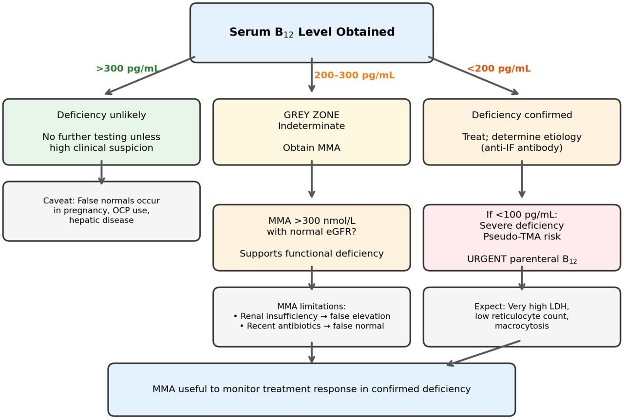
The serum cobalamin assay has well-established limitations. Levels above 300 pg/mL generally exclude deficiency; levels below 200 pg/mL generally confirm it.4 The intervening range represents genuine diagnostic uncertainty. Pregnancy, oral contraceptive use, HIV infection, and hepatic disease can all produce falsely normal or falsely low serum cobalamin independent of true tissue stores.3 Figure 1 presents a clinical algorithm for the interpretation of serum B12 and the role of confirmatory MMA testing.
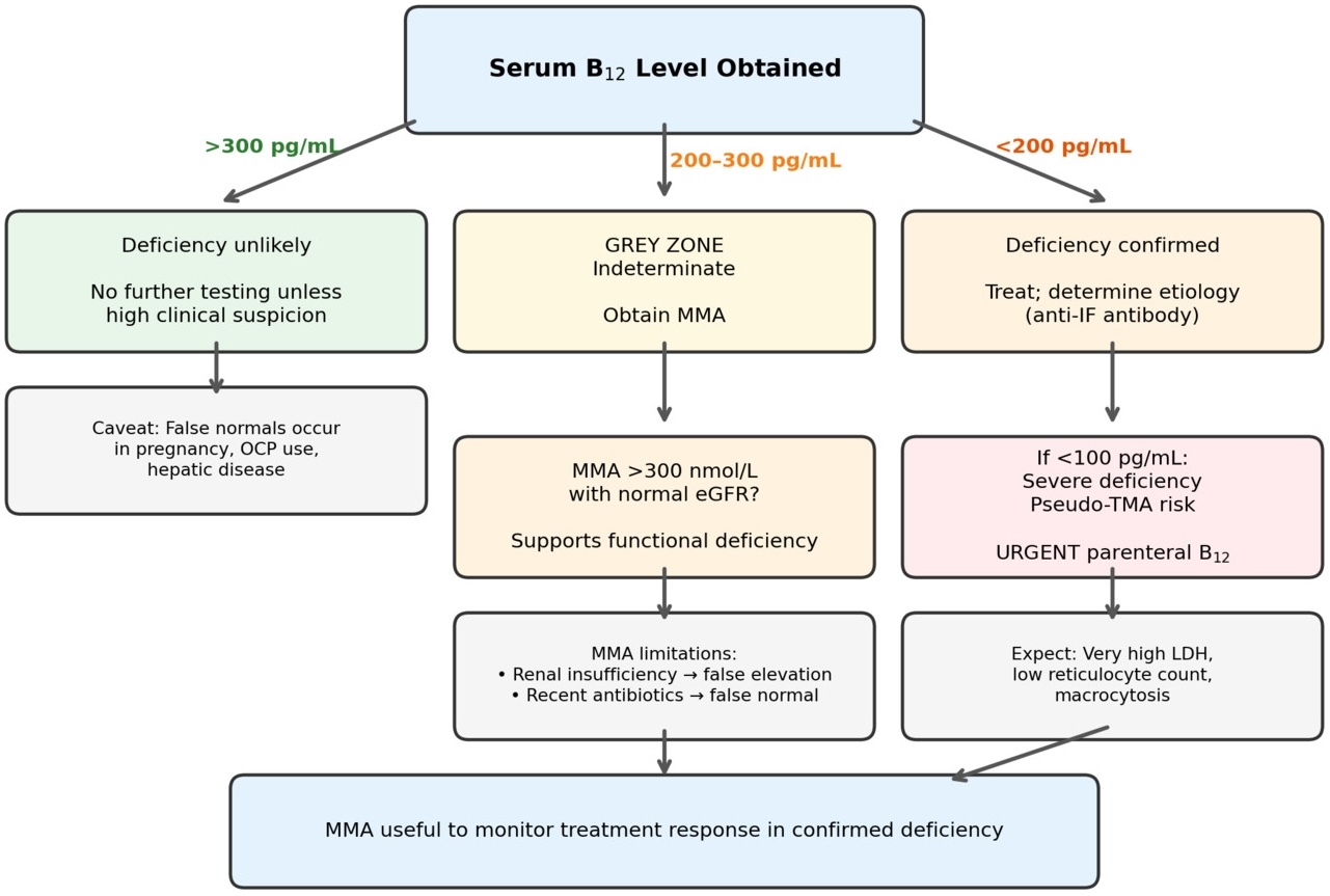
MMA is the most specific functional biomarker of intracellular B12 status. Cobalamin in its adenosylcobalamin form serves as a cofactor for methylmalonyl-CoA mutase, the mitochondrial enzyme that converts methylmalonyl-CoA to succinyl-CoA. When intracellular B12 is insufficient, this reaction fails, and MMA accumulates, often before serum cobalamin falls below 200 pg/mL.5
Clinicians should be aware of MMA limitations. Renal insufficiency elevates MMA independently of B12 status. Recent antibiotic use can produce falsely normal MMA by suppressing gut propionate-producing bacteria.4
Pseudo-TMA: Mechanism and Differentiation
Pseudo-TMA arises from intramedullary destruction of megaloblastic erythroid precursors.6 The marrow cannot make red cells properly. What follows looks exactly like TTP: hemolytic anemia, thrombocytopenia, schistocytes on smear, and an LDH that can exceed several thousand. The reticulocyte count is what separates the two conditions. In pseudo-TMA, it stays low because the marrow itself is the problem. In TTP, the marrow works fine, and the reticulocyte count rises in response to peripheral destruction.6,7
Table 1 summarizes the bedside differentiation. Our patient’s LDH exceeding 6,000 U/L, MCV of 115.5 fL, and reticulocyte count of 1.63% were collectively diagnostic of pseudo-TMA. However, evolving schistocytosis on serial smears raised concern for TTP. TTP occurs at approximately three per million per year but occupies a disproportionate place in clinical teaching, which inflates its perceived probability.7
Artifactual Bicarbonate Elevation
The chemistry panel reported serum bicarbonate greater than 45 mmol/L. This was wrong. When LDH reaches several thousand U/L, it consumes NADH in a side reaction within the enzymatic CO2 assay, and the analyzer reads that consumption as bicarbonate.10 We confirmed the interference on hospital day 3 when arterial blood gas showed a calculated bicarbonate of 23.4 mmol/L. The patient did not have metabolic alkalosis.
The spurious bicarbonate set the trajectory of the admission. The team anchored on metabolic alkalosis. Nephrology was consulted. An extensive workup was initiated. The fact that the patient had been vomiting made contraction alkalosis seem plausible, and nobody questioned the chemistry panel until the ABG came back two days later.
Cognitive Biases
Two biases are worth naming. The first is anchoring. Once the team accepted the bicarbonate as real, the workup followed that assumption and closed off other possibilities. Graber et al. showed that premature closure is the most common cognitive contributor to diagnostic error.11 The second is availability. TTP is rare, about three cases per million per year, but it looms large in clinical training.7 When schistocytes appeared on the smear, TTP jumped to the front of the differential, even though pseudo-TMA from B12 deficiency was far more likely given the clinical picture.
Polyautoimmunity and the Case for Screening
This patient’s constellation of pernicious anemia, T1DM, and vitiligo exemplifies polyautoimmunity. Among patients with T1DM, 15–20% harbor parietal cell antibodies and 2–6% develop overt pernicious anemia.8 Had annual B12 screening with reflex MMA testing been performed, the diagnosis would likely have preceded hematologic crisis.
Screening makes economic sense. Serum B12 and MMA are inexpensive relative to the downstream costs of missed diagnosis. This patient’s five-day hospitalization with subspecialty consultations and transfusions illustrates the disparity. Elevated MMA also predicts mortality independently of B12 level, which suggests a benefit from treating subclinical deficiency.12
These principles apply with particular urgency to patients on GLP-1 receptor agonist therapy. These medications can produce substantial caloric reductions.9 At intakes below 1,200 kcal/day in females or 1,800 kcal/day in males, micronutrient deficiencies become likely.9
Clinical Implications
The clearest takeaway is that serum cobalamin alone is not enough. Grey-zone levels need reflex MMA testing, and most primary care physicians are not ordering it. The cognitive errors in this case were not exotic. Anchoring and premature closure happen on every hospital service, and the only defense is knowing they will happen. GLP-1 receptor agonists are putting more patients at risk for nutritional deficiency every year, so B12 grey-zone interpretation is no longer a niche problem. Finally, patients with polyautoimmune clusters, especially T1DM, vitiligo, or autoimmune thyroiditis, deserve routine B12 screening. The test costs less than a copay. The hospitalization it prevents does not.
AI Disclosure
The authors used OpenEvidence, DoximityGPT, and UpToDate AI for literature search, clinical reference, literature organization, and citation formatting. Grammarly AI was used for grammar and sentence structure refinement. All content was critically reviewed and verified by the authors against primary sources. The authors retain full responsibility for the scientific content and clinical conclusions of this manuscript.