Brief Background
Rasmussen encephalitis (RE), also called Rasmussen’s syndrome, is a rare, chronic, and progressive neurological disorder thought to occur in two to four out of every 10,000,000 people.1 Unilateral hemispheric inflammation, refractory epilepsy, progressive neurological impairments, and cognitive impairment are features of the condition.2 Primarily, RE occurs in children aged two to 10, but adult cases, although very uncommon, are more difficult to diagnose and treat.1 Pathophysiologically, the exact cause of the disorder is unclear, but it can be attributed to an autoimmune response or a virus.2 At present, there are no cures for RE, and patients receive treatment with antiseizure medications (ASMs), immunomodulatory therapy, and surgery in pediatric refractory cases.2 In adults, however, RE has a slow onset and is complicated by the patient’s underlying psychiatric and substance abuse disorders, which are common in primary care practice.1,3
This case is particularly relevant to family medicine practitioners, as patients with RE may first present to a primary care physician with longstanding seizure disorders and behavioral health comorbidities before specialized neurological evaluation occurs. Timely recognition and appropriate referral are essential components of primary care management in such cases.
Case Report
A 42-year-old male with childhood-onset seizures, bipolar disorder, and polysubstance abuse came to medical attention due to a witnessed seizure episode. The patient reported that the previous day’s dose of Keppra (levetiracetam) was not taken. He denied any acute drug ingestion at the time of admission but admitted to drinking alcohol daily and crack cocaine of value $300 on a nightly basis for some days.
On physical exam, the patient was observed to be awake but confused with orientation to person, place, and time. His vital signs were blood pressure (BP) 147/93 mmHg (millimeters of mercury), heart rate (HR) 75/min, and RR 18/min. The neurological assessment showed altered mental status, right upper extremity (RUE) weakness, and right-sided tremor. In a psychiatric evaluation, the patient was found to have flight of ideas with no suicidal thoughts. His urine toxicology screen came back negative on admission to the hospital, while his blood alcohol level was less than 3.0 mg/dL (milligrams per deciliter). His complete blood count (CBC) results revealed a white blood count (WBC) of 10.0 x 10^9/L, hemoglobin (Hb) of 16.2 g/dL, and platelet count of 341 x 10^9/L. His creatinine level was 0.7 mg/dL, while other metabolic panels were within normal range.
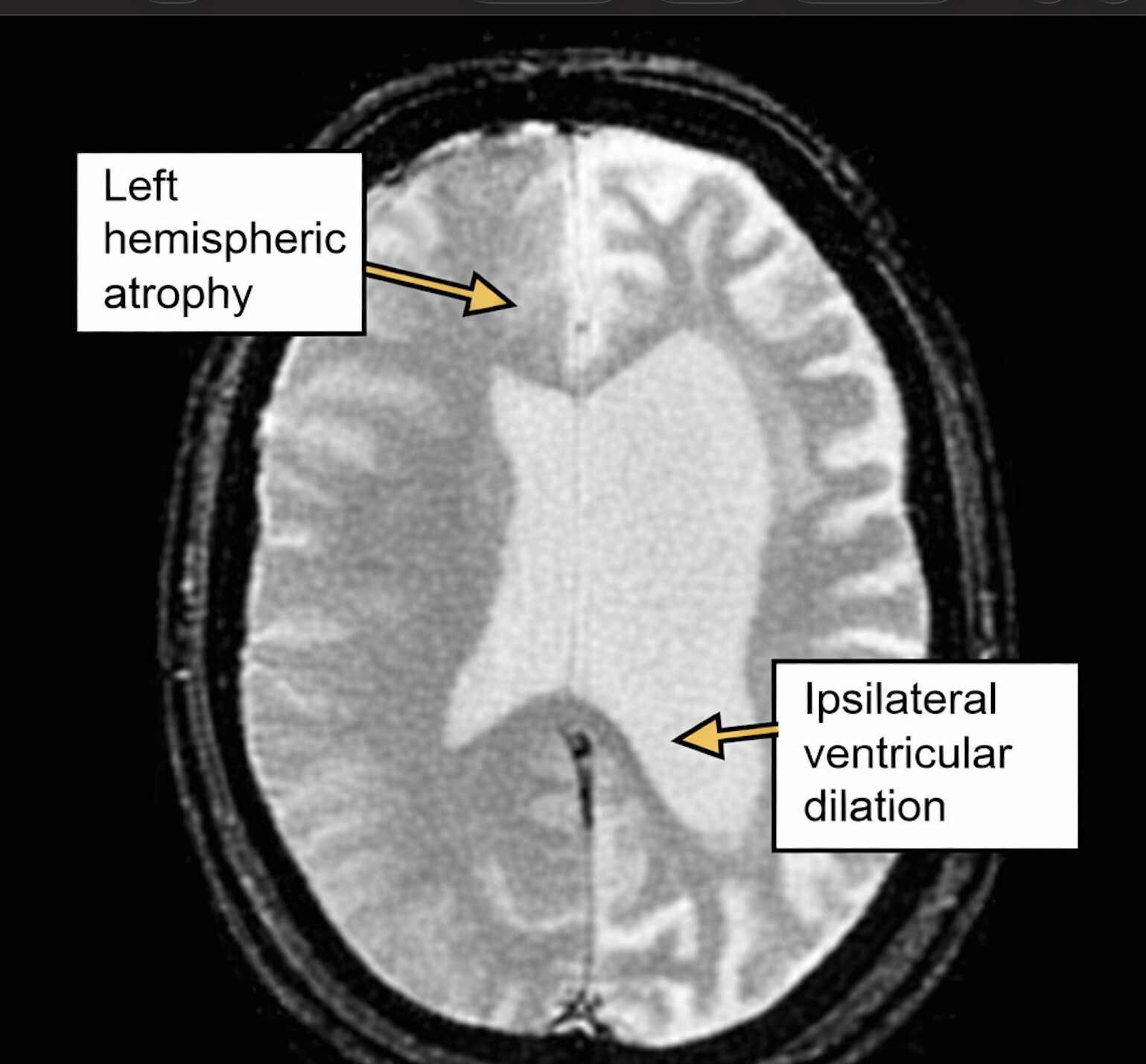
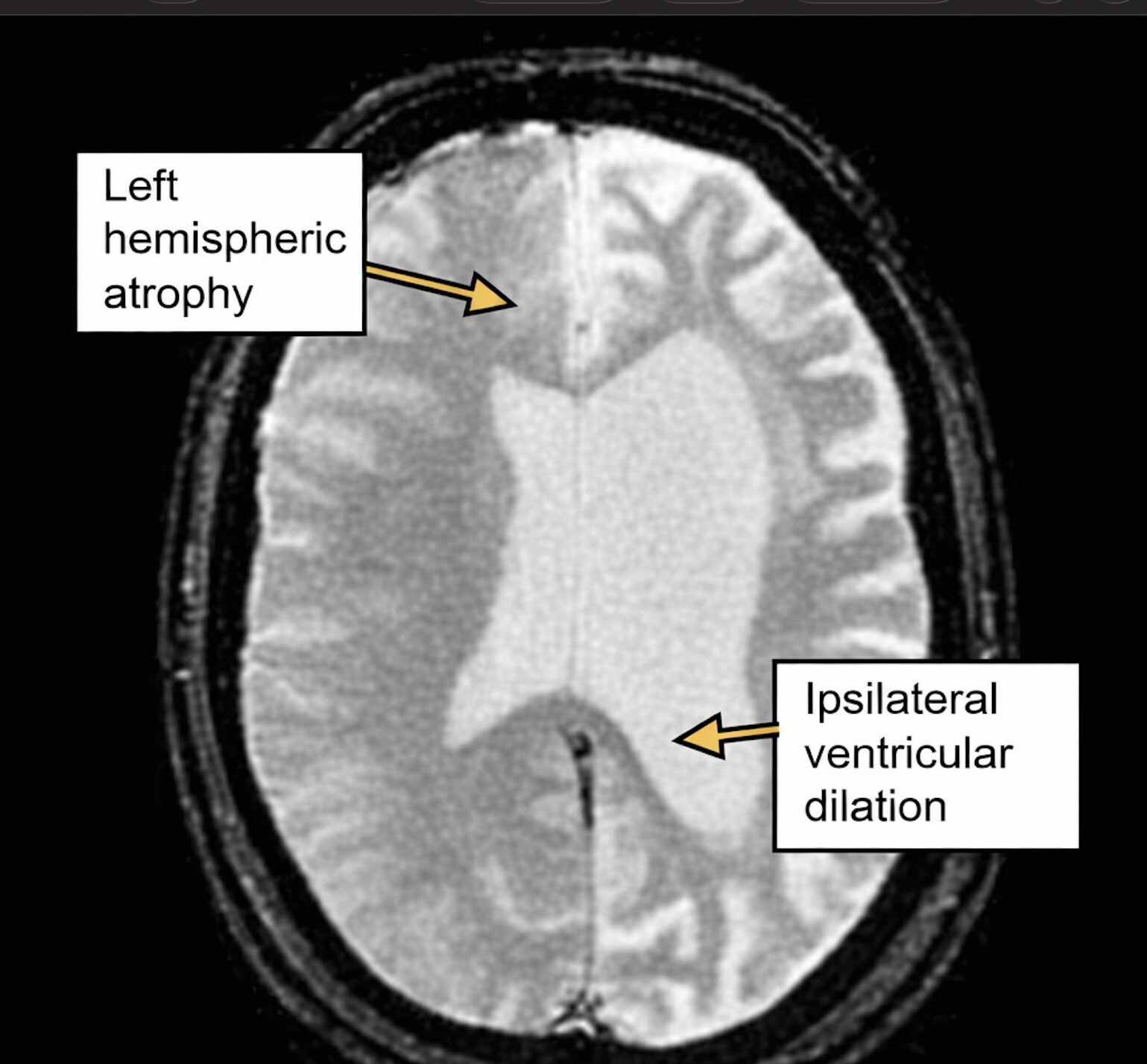
Computed tomography (CT) of the head without contrast showed no acute intracranial hemorrhage but revealed involutional changes in the left cerebrum and right cerebellum, ipsilateral dilation of the left lateral ventricle, and deep white matter low-attenuation changes in the left cerebral hemisphere. Levetiracetam 1,000 mg intravenously was administered in the emergency department, and the patient was admitted for further evaluation.
Brain magnetic resonance imaging (MRI) study performed on hospital day two showed diffuse encephalomalacia of the left cerebellar hemisphere, gliosis of the left frontal lobe, and atrophy of the right cerebellar hemisphere, consistent with crossed cerebellar diaschisis. Radiology interpretation: “These findings can be seen as consequences of previous hemispheric infarct or Rasmussen encephalitis.” CT angiography (CTA) of the head and neck scan demonstrated decreased left A3 and M3 runoff, mild left cerebral atrophy with enlargement of the left lateral ventricle, and mild decrease in right cerebellar hemisphere runoff.
Electroencephalography (EEG) showed left-hemispheric slowing, no posterior dominant rhythm, and asymmetric background activity in more than 50% of the time, without any ictal activity.
These results are shown in Figure 1 below.
Consultations with neurology and psychiatry were made, and the patient was placed on levetiracetam 1,000 mg intravenous administration twice a day and oxcarbazepine (Trileptal) 300 mg oral administration twice a day. Lorazepam (Ativan) 1 mg intravenous administration was available as needed for breakthrough seizures. The Clinical Institute Withdrawal Assessment for Alcohol (CIWA) protocol for alcohol withdrawal management was done by administering thiamine 100 mg intravenously once a day, folic acid 1 mg oral administration once a day, and multivitamin supplements. Olanzapine (Zyprexa) and ziprasidone (Geodon) were initiated following the recommendation of a psychiatrist. The speech therapist revised the dietary intake to thin liquids mixed with crushed medications and the chin-tuck technique. Deep vein thrombosis (DVT) prophylaxis continued with enoxaparin sodium (Lovenox) 40 mg, subcutaneously once daily.
Discussion
This case presents a diagnostically challenging adult presentation of RE in the setting of longstanding polysubstance use disorder, bipolar disorder, and prior ischemic stroke—all of which can independently produce neuroimaging abnormalities and refractory epilepsy. Adult-onset RE is rare, with fewer than 100 cases described in the literature, and it is characterized by a more prolonged prodromal phase and slower hemispheric atrophy compared with childhood-onset RE.1,3 The neuroimaging constellation of diffuse left cerebellar encephalomalacia, focal frontal gliosis, and crossed cerebellar diaschisis (Figure 1) is consistent with the unilateral hemispheric degeneration that defines RE.2
Diagnostic challenges were greatly increased by the concomitant presence of a substance abuse disorder. Long-term usage of both cocaine and alcohol can, on their own, lead to cerebrovascular damage, alterations in the white matter, and a decreased seizure threshold. All of these factors complicate the neuroimaging picture.4 Within the realm of general practice medicine, adults with childhood epilepsy, along with comorbid psychiatric illness, are generally under long-term care before neurology consultation. The combination of unilateral cerebral atrophy, unilateral cerebral slowing, and seizures while maintaining sufficient levels of antiepileptic drugs necessitates suspicion of RE.
The EEG findings of focal left hemispheric slowing without posterior dominant rhythm are consistent with the electrographic dysfunction seen in RE, where hemispheric slowing and focal epileptiform discharges are characteristic.3 While definitive diagnosis requires brain biopsy demonstrating T-lymphocyte infiltration and microglial nodules, the clinical and radiological constellation in this patient met high-suspicion criteria per European Consensus Statement diagnostic guidelines.4
Management of the adult-onset RE condition is still difficult because there are no evidence-based guidelines yet for adults with the disease. Immunotherapy is effective in pediatric patients; however, its efficacy in adults remains debatable.4 Levetiracetam and oxcarbazepine were used in the treatment of seizures associated with RE.1 The use of various specialists from different fields, such as neurology, psychiatry, internal medicine, and speech therapists, proved to be critical in this case and highlighted the importance of family physicians’ contribution in providing coordination among specialists.2 There is a strong possibility that lack of a primary care physician has been a factor behind delayed diagnosis and treatment, as well as poor adherence to prescribed medication.
The described patient’s case highlights RE as a differential in adults with complicated clinical presentation of seizure disorder in the setting of drug addiction and mental disorders. Primary care providers must recognize the importance of their role in the diagnostic work needed for the early detection of rare disorders which can improve the outcome.